

Introduction
Chicken egg white has a rich protein composition and functional properties, which are widely used in the food, pharmaceutical, and nutraceutical industries (Ji et al., 2020). It consists of approximately 88% of water, 0.2% of fat, and 0.8% of ash, and 11% of protein, with the protein fraction comprising several bioactive components that contribute to its technological and biological significance (Campbell et al., 2003). Among them, lysozyme and ovomucin are of particular interest because of their antibacterial, antiviral, and antigenic properties (Stadelman et al., 2017).
Lysozyme is a low-molecular-weight enzyme (14.4 kDa) and constitutes approximately 3.4% of the total egg white proteins (Abeyrathne et al., 2014). It is well known for its excellent bacteriostatic, bactericidal, and antiviral properties, primarily due to its ability to hydrolyze the β-(1,4)-glycosidic linkages in bacterial cell walls that can be used as an active ingredient in food preservation, pharmaceuticals, and medical applications (Abeyrathne et al., 2013; Silvetti et al., 2017). Ovomucin is another important protein in the egg white. Chicken egg white contains 3.5% of ovomucin, and its molecular weight is 5.5–8.3×10³ kDa (Abeyrathne et al., 2014). It is a glycoprotein consisting of α- and β-subunits that are bound by disulfide bonds (Li et al., 2022; Stadelman et al., 2017). The two subunits create a complex structure that is responsible for its functional properties, such as the formation of various gels and maintaining the structural stability of the egg white matrix (Hiidenhovi, 2015). Also, it has antibacterial, antiviral, antitumor properties, and anti-hemagglutination activity against the influenza virus. Furthermore, its excellent water-binding, emulsifying, and foaming properties make it highly relevant for food processing applications.
Despite their importance, the efficient separation and purification of lysozyme and ovomucin remain challenging. Conventional methods such as ion-exchange chromatography, membrane filtration, and precipitation techniques have been widely employed (Abeyrathne et al., 2013). Ion-exchange chromatography can achieve high purity levels (>90%), but typically requires multiple steps, expensive resins, and high operational costs, limiting its scalability (Kisley, 2015). Membrane-based separation methods provide moderate selectivity; however, they are often affected by membrane fouling and require significant energy input (AlSawaftah et al., 2021). Precipitation-based approaches are relatively simple and cost-effective but suffer from low selectivity and co-precipitation of impurities, particularly in the case of ovomucin (Li et al., 2022).
Previously reported lysozyme extraction methods generally achieve yields of 20%–60% with purity ranging from 70%–95%, depending on process complexity, whereas ovomucin recovery via precipitation methods typically ranges from 30%–60% with relatively low purity due to aggregation and co-precipitation of other egg white proteins (Abeyrathne et al., 2013). In the present study, the separation method will reduce reliance on multi-step chromatographic procedures and limits chemical usage, which may help decrease operational complexity and improve its potential scalable applications compare with the previous reported methods.
The present method, foam-based separation, is a surface-driven technique in which proteins are selectively adsorbed at the gas-liquid interface during bubble formation. Proteins with higher surface activity, which are influenced by their physicochemical properties such as hydrophobicity, charge destruction, and structural flexibility, preferentially migrate to and stabilize the air-water interface. As a result, these proteins become enriched in the foam phase, while proteins with higher solubility and lower surface activity remain in the bulk liquid phase (Noble et al., 1998; Sochacki et al., 2025; Sunkesula et al., 2020). In egg white systems, ovomucin, a high-molecular-weight fibrous glycoprotein, plays a major role in foam stabilization through its structural characteristics and intermolecular interactions, thereby enriching the foam fraction. In contrast, lysozyme is a smaller, highly soluble, and positively charged globular protein that tends to remain in the aqueous phase. This difference in interfacial behavior provides the basis for selective separation using foam formation as an initial fractionation step (Beveridge, 1973).
Based on these considerations, the present study aims to develop a simple, cost-effective, and scalable foam-based method for the sequential isolation of lysozyme and ovomucin from chicken egg white. This study provides a practical alternative strategy for the recovery of high-value egg white proteins with potential applications in food and bioprocessing industries.
Materials and Methods
Fresh large-sized chicken eggs were purchased from a local market in Badulla, Sri Lanka, and used within 24 h of purchase. Egg freshness was verified prior to use by candling. After selecting fresh eggs, they were stored at 4°C until analysis to minimize protein degradation. Egg whites were carefully separated from yolks and processed individually, without pooling, as only fresh eggs of consistent quality were selected, thereby minimizing variability among samples. Amberlite FPC 3500 cation-exchange resin (styrene-divinylbenzene, total exchange capacity ≥2.6 eq/L, H+-form) was obtained from Acros Organics. Chemicals (NaCl, Citric acid, Acetic acid, Sodium dodecyl sulfate) were purchased from Sigma-Aldrich (St. Louis, MO, USA). All chemicals used were of analytical grade.
All experiments were conducted in triplicate (n=3). A schematic flow of the process is presented in Fig. 1. The overall experimental procedure consisted of four main steps: (i) foam generation and fractionation, (ii) optimization of foam redissolution, (iii) lysozyme separation by cation-exchange adsorption, and (iv) ovomucin recovery by isoelectric precipitation.
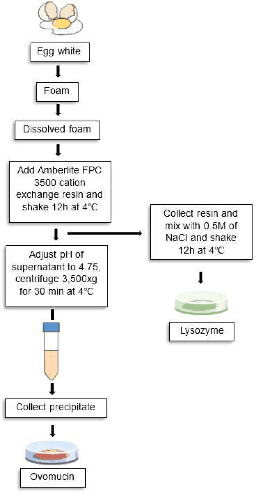
Fresh, high-quality eggs were cleaned by wiping and allowed to air-dry before processing (Kudre et al., 2018). The egg whites were carefully separated from the yolks and subjected to foam generation using a hand whipper operated at constant speed for 5 min at room temperature (25°C). No external airflow was applied during the foaming process. After whipping, the foam was allowed to stand undisturbed for 5 min to facilitate phase separation. After a 2 h rest at 4°C, the residual liquid was re-whipped under the same conditions to generate additional foam. This procedure was repeated three times to maximize foam recovery. All foam fractions were stored at 4°C overnight prior to further processing.
To evaluate the efficiency of foam redissolution, different agents such as NaCl, citric acid, acetic acid, and sodium dodecyl sulfate (SDS) were used. Solutions of NaCl, citric acid, and acetic acid (1 M) were prepared using distilled water. One gram (1 g) of egg white foam was then added separately to each solution and homogenized to promote protein redissolution. For SDS treatment, solutions containing 0.4%, 0.5%, 0.6%, and 0.7% (w/v) SDS were prepared. The pH of each SDS solution was adjusted to 5.0, 7.0, or 9.0, and verified with a calibrated pH meter (Model HI5221-02, Hanna Instruments, Woonsocket, RI, USA). After pH adjustment, 1 g of egg white foam was added to the solution and homogenized thoroughly. The extent and rate of redissolution were assessed by visual observation of foam breakdown and the formation of a homogeneous solution. The condition showing complete dissolution was selected for subsequent experiments.
Lysozyme was isolated from the redissolved foam solution with slight modifications from a previously reported method (Abeyrathne et al., 2014). Briefly, Amberlite FPC 3500 cation-exchange resin was suspended in 500 mL of distilled water and equilibrated to pH 11.7 by gradual addition of 1 N NaOH under continuous stirring. After equilibration, the excess water was removed. The conditioned resin was then mixed with the dissolved foam solution to allow selective adsorption of lysozyme. The mixture was gently stirred using an overhead stirrer at the lowest speed for 12 h in a cold room maintained at 4°C. After incubation, the resin was separated from the mixture for lysozyme recovery. To elute bound lysozyme, the resin was treated with two volumes of 0.5 M NaCl, pH 11.7, and stirred for 12 h at 4°C. The supernatant containing the released lysozyme was collected. This elution step was repeated twice to ensure maximum recovery. All collected fractions were pooled, concentrated, and desalted by ultrafiltration, followed by freeze-drying using a lyophilizer (Model 05512, iLShinBioBase, Dongducheon, Korea).
The solution after lysozyme removal was used for ovomucin separation according to the method described by (Abeyrathne et al., 2014). The pH of the solution was adjusted to 4.75 to induce isoelectric precipitation of ovomucin. The mixture was then centrifuged at 3,400×g for 30 min at 4°C. The resulting precipitate was collected and washed twice with cold distilled water 9pH adjusted to 4.75) to remove loosely bound impurities and co-precipitated proteins. After washing, the precipitate was re-suspended and centrifuged under the same conditions to improve purity. The final precipitate containing crude ovomucin was collected, concentrated, and further dried for subsequent analysis.
Protein profiles of the separated fractions were analyzed using sodium dodecyl sulfate–polyacrylamide gel electrophoresis (SDS-PAGE) with a 10% polyacrylamide gel (Bio-Rad Mini-PROTEIN Tetra System, Shanghai, China). Protein bands were visualized after staining, and molecular weight markers were used for comparison.
Tandem mass spectrometry was performed to confirm the identity of the separated proteins. For MALDI-TOF-MS/MS (matrix-assisted laser desorption/ionization—time-of-flight) analysis, the protein bands corresponding to ovomucin and lysozyme were excised from the SDS-PAGE gel and subjected to in-gel digestion with trypsin at 37°C overnight. Following digestion, the peptide extracts were recovered by centrifugation at 10,000×g for 3 min. One microliter of the desalted tryptic digest was mixed with 0.6 μL of a supersaturated α-cyano-4-hydroxycinnamic acid (CHCA) matrix solution prepared in 50% acetonitrile (ACN) containing 0.1% trifluoroacetic acid (TFA), and the mixture was spotted onto a MALDI target plate. Samples were analyzed using a matrix-assisted laser desorption/ionization time-of-flight mass spectrophotometer (5800 MALDI-TOF/TOF, AB SCIEX). The instrument was equipped with an Nd: YAG laser operating at a wavelength of 349 nm. The acceleration voltage was set at 2 kV, and the collision energy was maintained at 2 kV. Spectra were acquired in positive ion mode under automatic data acquisition settings. The MS scan range was 800–4,000 Da. Precursor ions with a signal-to-noise ratio greater than 50 were selected for MS/MS analysis. For each sample spot, ten precursor ions were chosen, and MS/MS spectra were accumulated 2,500 times. Protein identification was performed by searching the acquired spectra against the NCBI database using Mascot software (Version 2.2), and the final protein assignments were based on the best matching scores.
The weights of lysozyme and ovomucin were determined using a digital balance (Pioneer PX124KR, OHAUS Corporation, Parsippany, NJ, USA) following freeze-drying and subsequent grinding into powder form.
The purity of each separated protein was calculated by converting the density of protein bands in the gel picture to the percent of the total gel density (Eq. (1); Abeyrathne et al., 2014). The yields of lysozyme and ovomucin were calculated using their theoretical values in egg white. Yield was calculated with the ratio between lyophilized protein and the theoretical amount presented (Eq. (2); Omana and Wu, 2009).
Fourier transform infrared (FTIR) spectroscopy (ALPHA, Bruker Optics, Ettlingen, Germany) was employed to analyze the structural characteristics of the separated proteins. FTIR operates based on the interference of two infrared beams, generating an interferogram that is mathematically transformed into an absorption spectrum (Tatulian, 2019). The technique measures the absorption of infrared radiation by the samples as a function of wavelength, and the resulting absorption bands provide information regarding molecular composition and structural features (Kong and Yu, 2007). In the present study, spectra were recorded in the range of 1,600–1,700 cm–1, corresponding to the amide I region, which is highly sensitive to protein secondary structure. Therefore, FTIR analysis was used to confirm the structural integrity of the separated proteins and to correlate their structural characteristics with functional properties, including solubility, emulsifying activity, foaming capacity, gelation behaviour, and structural stability. The functional attributes are strongly influenced by secondary structural elements, hydrogen bonding, hydrophobic interactions, and potential chemical modifications (Jabs, 2005).
All experiments were performed in triplicate, and the results are expressed as mean±SD. Statistical analysis was conducted using R statistical software (version 4.5.2). Differences between groups (lysozyme and ovomucin) were evaluated using an independent samples t-test. A p-value of less than 0.05 was considered to indicate statistical significance.
Results and Discussion
Efficient redissolution of egg white foam is essential for maximizing protein recovery. In this study, different agents, including NaCl, citric acid, acetic acid, and SDS, were evaluated for their ability to disrupt foam structure and solubilize proteins (Figs. 2A, B, C, and D).
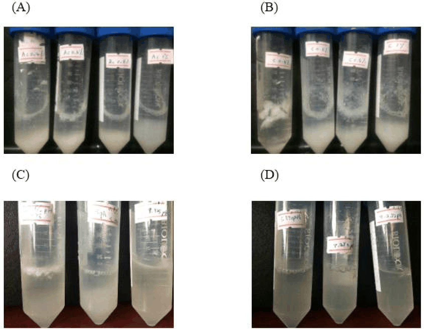
Acidic solutions (citric acid and acetic acid) showed partial foam destabilization; however, complete dissolution was not achieved. The reduced solubility of egg white foam under acidic conditions is largely driven by protein aggregation (Gomes and Pelegrine, 2012). As the pH drops toward the isoelectric point of major proteins like ovalbumin, their net charge nears neutrality. This loss of electrostatic repulsion allows the proteins to cluster and precipitate, which accounts for the higher sedimentation levels observed in the citric acid treatments (Shimoyamada et al., 2018). Compared to the effects of acetic acid, citric acid induced greater protein precipitation. This disparity is potentially due to the multivalent nature of the citrate ion, which, alongside its strong buffering capacity, enhances the cross-linking and subsequent aggregation of the egg white proteins (Brudar and Hribar-Lee, 2021; Wurm et al., 2020). Similarly, NaCl treatment did not effectively collapse the foam structure, suggesting that ionic strength alone is not adequate for complete protein solubilization. As ionic strength increases, the salt ions compete with protein functional groups for available water molecules, effectively stripping the hydration shell from the proteins. This dehydration promotes hydrophobic protein-protein interactions, leading to accelerated aggregation and precipitation (Wang et al., 2022).
In contrast, SDS demonstrated superior performance. Among the tested conditions, 0.6% SDS (20 mL) resulted in partial destabilization at pH 5 and pH 7, while complete dissolution of the foam was observed at pH 9. This can be explained by the amphiphilic nature of SDS, which disrupts hydrophobic interactions and enhances protein solubility (Jafari et al., 2018). However, higher concentrations of SDS may pose potential risks to human health (Boelhouwer et al., 2013). Among 0.5%, 0.6%, and 0.7% SDS concentrations, 0.6% SDS exhibited the highest redissolving efficiency. was selected as the optimal condition for subsequent separation processes. Consequently, 0.7% SDS was not further investigated, therefore, 0.6% SDS was selected as the optimal condition for subsequent separation processes.
In this study, a relatively low SDS concentration was used to facilitate foam redissolution. Previous studies have reported that SDS at low concentrations has minimal effect on lysozyme structure (Chodankar et al., 2008). Furthermore, the large molecular size and complex glycoprotein structure of ovomucin reduce its susceptibility to SDS-induced structural changes (Omana et al., 2010). Residual SDS present in the lysozyme and ovomucin fractions was subsequently removed by ultrafiltration during the purification process. The SDS-PAGE profiles revealed that the target proteins were successfully separated (Fig. 3). The lysozyme fraction showed a clear band around 14–16 kDa, which corresponded to its known molecular weight and indicated good purity. In contrast, the ovomucin fraction showed high-molecular-weight bands corresponding to its α-subunit, indicating its complex glycoprotein structure. Both fractions showed minimal contamination, confirming the efficacy of the separation technique. The intermediate fractions demonstrated the increasing elimination of proteins over the consecutive phases.
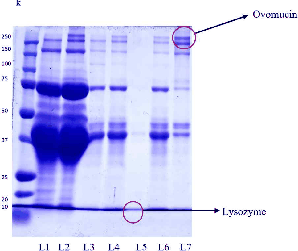
The identities of the isolated proteins were validated by MALDI-TOF-MS (Table 1). The observed peptide mass fingerprints were consistent with known lysozyme and ovomucin sequences from Gallus gallus. Lysozyme was found to have a molecular weight of around 16.2 kDa and an isoelectric point (pI) of 9.07. Ovomucin, the α-subunit, has a molecular weight of approximately 233.4 kDa and a pI of 5.6. These results confirm the reliability of the separation method and demonstrate that the proteins retained their molecular identity after processing.
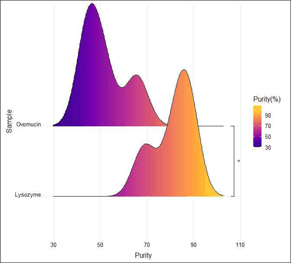
According to the results of the present study, the recovered lysozyme exhibited a final weight of 0.49±0.03 g, with a purity of 81.18±10.73% and an overall yield of 26.38±1.35% (Table 2). The purity level of lysozyme obtained in the present study was approximately 81%, indicating that the selected separation method was relatively efficient. Wu et al. (2015) stated that lysozyme purity typically ranges from about 70% to over 90%, depending on the purification strategy employed. Single-step purification methods generally achieve 70%–80% purity, whereas multi-step chromatographic procedures can exceed 90% purity, often at the expense of decreased overall yield (Wulandari et al., 2015). Therefore, the results of the present study suggest a balanced extraction approach, achieving moderately high purity while minimizing excessive protein loss.
| Protein | Weight (g) | Purity (%) | Yield (%) |
|---|---|---|---|
| Lysozyme | 0.49±0.03 | 81.18±10.73 | 26.38±1.35 |
| Ovomucin | 0.86±0.06 | 41.93±25.09 | 44.68±2.89 |
Lysozyme typically accounts for approximately 3%–3.5% of the total protein in egg white. Previous studies have reported extraction yields for egg white lysozyme generally ranging from 20%–30%. In the present study, the lysozyme yield was 26.38%, which is consistent with earlier findings (Chang et al., 2000). The extraction yield is strongly influenced by the purification strategy employed, including salt precipitation, ion-exchange chromatography, ultrafiltration, and affinity-based techniques (Shahmohammadi, 2018). For example, Hou and Lin (1997) reported yields of 57% and 59% using alcohol-insoluble solids and cross-linked alcohol-insoluble solids from sweet potato leaves, respectively, and 58% when applying a linear salt gradient. Du and Yang (2007) achieved an enzyme recovery rate of 87% using ion-exchange resin. In contrast, Qing-hong and Xiu-lian (2010) reported an extraction yield of 37% using an optimized salting-out method. These variations highlight the substantial impact of purification methodology on lysosome recovery efficiency.
The recovered ovomucin had a final weight of 0.86±0.06 g, with a yield of 44.68±2.86% and a purity of 41.93±25.09% (Table 2). The purity (41.91%) was relatively low and exhibited considerable variability (±25.09%). Similar observations have been reported in previous studies, where crude ovomucin fractions frequently contain substantial amounts of co-precipitated proteins, including ovalbumin, ovotransferrin, and other egg white constituents (Omana and Wu, 2009). Ovomucin is a high-molecular-weight glycoprotein complex composed of α- and β-subunits interconnected by disulfide bonds and extensively associated with carbohydrate units. Due to its gel-forming and fibrous nature, ovomucin readily forms aggregates and interacts with other proteins, thereby complicating selective purification and contributing to reduced purity levels (Beck et al., 2025).
However, the yield obtained in the present study falls within the range reported in previous investigations on egg white fractionation. Ovomucin typically accounts for approximately 1.5%–3.5% of the total egg white protein. However, reported extraction yields vary considerably (30%–60%) depending on the separation technique employed, including isoelectric precipitation, salt-aggregation, dilution-centrifugation methods, and sequential washing steps (Li et al., 2022). The relatively high recovery (44.68%) observed in this study suggests that the applied precipitation-based method was effective in isolating an ovomucin fraction (Wang et al., 2015).
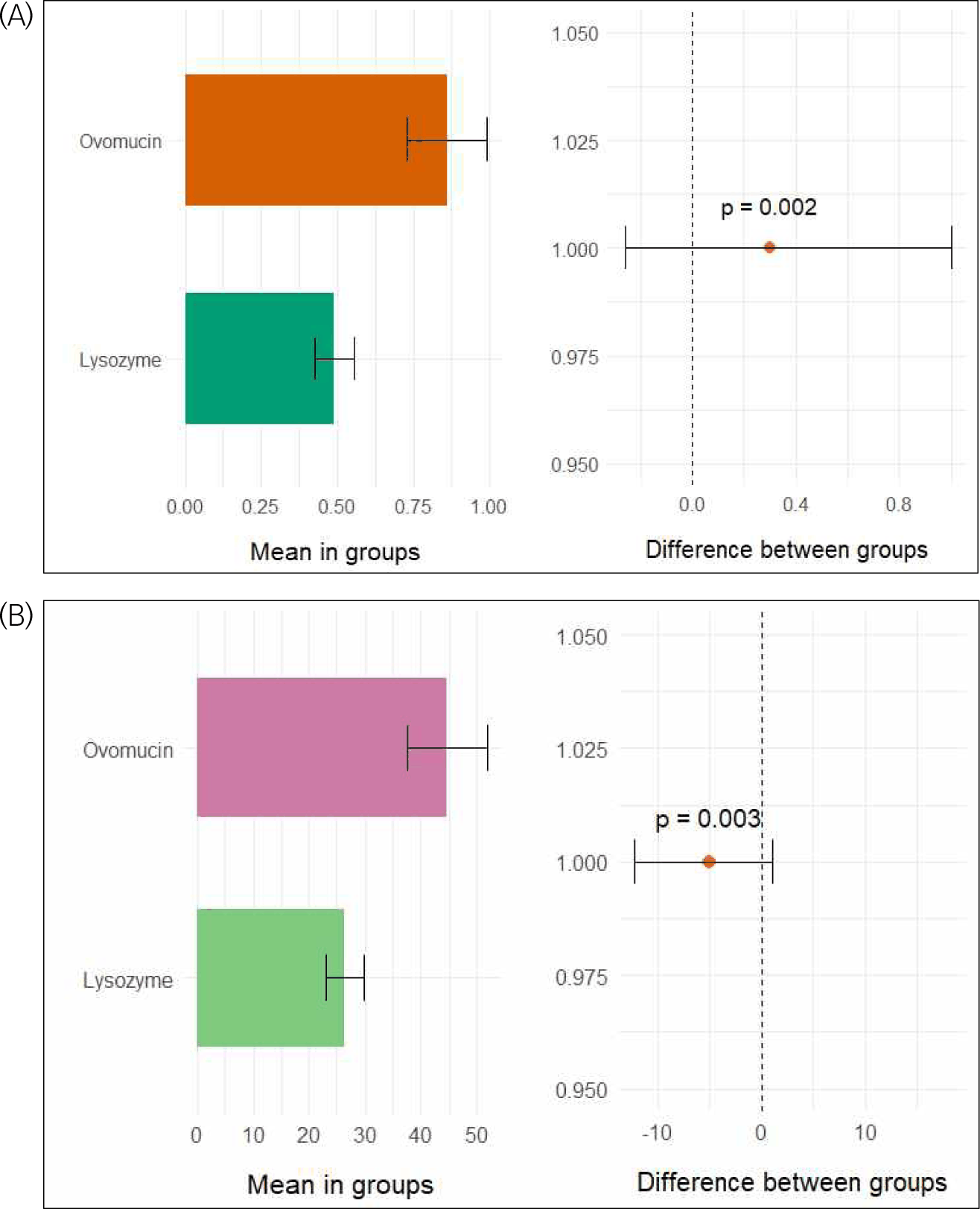
The weight, yield, and purity of lysozyme and ovomucin were evaluated to determine the effectiveness of the described approach. Significant differences were found between the two proteins (p<0.05). Ovomucin had a substantially greater recoverable weight than lysozyme (Fig. 4A). This finding is primarily attributed to the aggregation behavior of ovomucin and its tendency to co-precipitate with other egg white proteins near its isoelectric point, which contributes to increased mass recovery during precipitation. Similarly, ovomucin had a substantially higher yield than lysozyme (Fig. 4B). The higher yield of ovomucin can be attributable to its efficient recovery by isoelectric precipitation, whereas lysozyme’s lower yield may be due to inadequate adsorption to the cation-exchange resin or losses during elution and processing. In contrast, lysozyme had much higher purity than ovomucin (Fig. 5). These findings suggest a trade-off between purity and yield, which can be explained by differences in charge interactions, solubility, and protein structural properties. Lysozyme, being positively charged at alkaline pH, shows strong and selective binding to cation-exchange resin, resulting in higher purity but lower yield due to incomplete recovery during adsorption and elution. In contrast, ovomucin, a high-molecular-weight fibrous glycoprotein with limited solubility near its isoelectric point, undergoes bulk precipitation along with other proteins, leading to a higher yield but reduced purity.
Overall, the developed extraction protocol achieved a balanced performance, providing satisfactory recovery and purity levels comparable to previously reported methods. However, the foam-based fractionation strategy relies exclusively on proteins partitioning into the foam phase during whipping; consequently, incomplete transfer of lysozyme into the foam fraction likely contributes to the moderate yield observed. Similarly, although the recovery of ovomucin was relatively high, its purity remained limited due to the intrinsic complexity of the foam-derived protein matrix. Specifically, co-existing foam-active proteins such as ovalbumin and ovotransferrin may remain associated with the ovomucin network through intermolecular interactions, leading to co-precipitation during isoelectric precipitation. These factors collectively reduce purification efficiency. Therefore, further optimization—such as precise pH and ionic strength control, additional washing steps, and incorporation of chromatographic polishing—may enhance purity without substantially compromising yield.
The observed trade-off between purity and yield is mainly due to differences in charge solubility and protein structural properties (Che Hussian and Leong, 2024). Lysozyme, with a high isoelectric point, carries a strong positive charge at alkaline pH and selectively binds to a cation-exchange resin, resulting in higher purity but lower yield due to incomplete recovery (Coskun, 2016). In contrast, ovomucin, a high-molecular-weight glycoprotein, has low solubility near its isoelectric point, leading to aggregation and co-precipitation with other proteins, thereby increasing yield but reducing purity (Abeyrathne et al., 2014). Additionally, the temporary use of SDS during foam redissolution may enhance protein solubility by disrupting hydrophobic interactions; however, since it was used at low concentration and removed during purification, its overall impact on separation efficiency is limited (Chodankar et al., 2008; Omana et al., 2010).
FTIR spectroscopy was employed to confirm the structural characteristics and functional groups of the separated lysozyme and ovomucin fractions (Figs. 6A and B). Both fractions exhibited characteristic protein absorption bands, including prominent signals in the amide I (approximately 1,600–1,700 cm–1) and amide II (approximately 1,500–1,600 cm–1) regions, corresponding to C=O stretching and N-H bending vibrations of the peptide backbone, respectively (Kumar et al., 2014). The presence of these typical proteins indicates that the fundamental structural features of lysozyme and ovomucin were preserved during the separation process, thereby confirming their successful isolation.
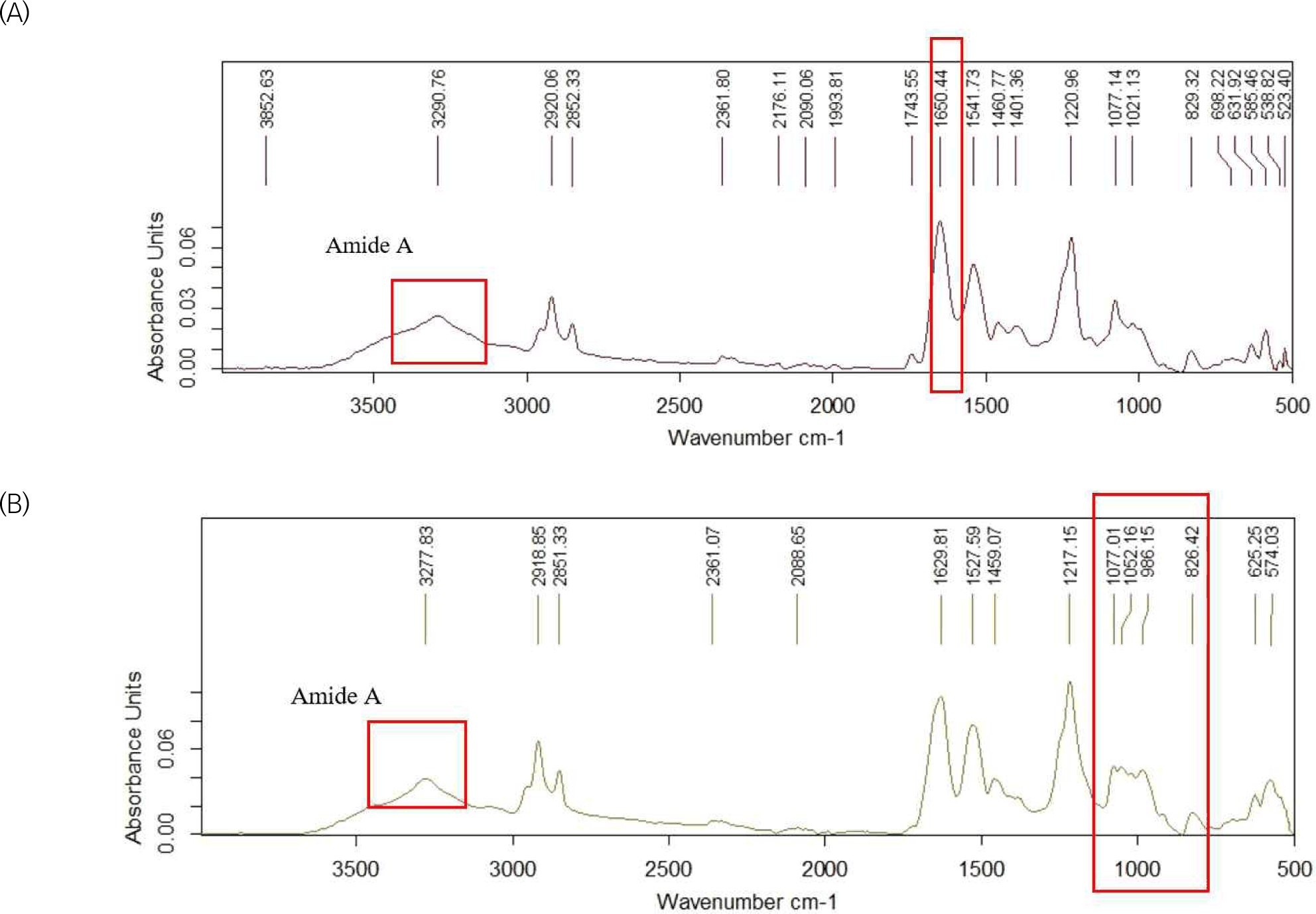
The FTIR spectrum of the lysozyme (Fig. 6A) fraction exhibited a broad absorption band at 3,290.76 cm–1, attributed to N-H stretching vibrations (amide A) and hydrogen bond O-H groups (Tonan and Ikawa, 1996). The bands at 2,920.06 cm–1 and 2,852.33 cm–1 correspond to asymmetric and symmetric C-H stretching vibrations of aliphatic residues (Urbanová et al., 2026). A strong absorption peak observed at 1,650.44 cm–1 represents the amide I region, primarily associated with C=O stretching of the peptide backbone. The position of this band near 1,650 cm–1 indicates a predominance of α-helical structure, which is consistent with previously reported FTIR spectra of native lysozyme from egg white (Arunkumar et al., 2019). The band at 1541.73 cm–1 corresponds to the amide II region (N-H bending coupled with C-N stretching), further confirming the preservation of the peptide backbone. Additional peaks at 1,460.77, 1,401.36, 1,220.96, 1,077.14, and 1,021.13 cm–1 are attributed to C-N, C-O, and C-H bending vibrations. The presence of well-defined amide I and II bands without significant peak shifts suggests that the extraction process did not markedly alter the native secondary structure of lysozyme, in agreement with previous studies on isolated egg white proteins (Balan et al., 2019).
FTIR spectrum of the ovomucin (Fig. 6B) fraction exhibited a broad absorption band at 3,277.83 cm––1, corresponding to overlapping O-H and N-H stretching vibrations. The relatively strong intensity of this band reflects extensive hydrogen bonding, which is typical of glycoproteins and has been reported previously for egg white ovomucin. The C-H stretching vibrations observed at 2,918.85 cm–1 and 2,851.33 cm–1 are associated with aliphatic groups (Pasquini, 2020). The amide I band appeared at 1,629.81 cm–1, while the amide II band was observed at 1,527.59 cm–1, corresponding to C=O stretching and N-H bending/C-N stretching vibrations of the peptide backbone, relatively. The amide I band near 1,630 cm–1 suggests a higher β-sheet content compared to lysozyme, which agrees with previously reported structural characteristics of ovomucin in the studies (Hu et al., 2006). A distinct feature of the spectrum was the strong absorption in the 1,200–1,000 cm–1 region, particularly at 1,217.15, 1,077.01, 1,052.16, 968.15, and 826.42 cm–1, attributed to C-O-C and C-O stretching vibrations of carbohydrate units. Similar carbohydrate-associated bands have been consistently reported for ovomucin due to its glycoprotein nature (Invernizzi et al., 2018). Overall, the spectral profile is consistent with previous findings and indicates that the separation process preserved the characteristic structural features of ovomucin.
Conclusion
This study developed a foam-based approach for the sequential isolation of lysozyme and ovomucin from chicken egg white. The method enabled the separation of both proteins using a combination of foam fractionation, cation-exchange adsorption, and isoelectric precipitation. The obtained lysozyme fraction showed relatively high purity, while the ovomucin fraction exhibited higher recovery but lower purity, indicating a trade-off between selectivity and yield. Structural analyses suggested that the main protein characteristics were largely preserved during the process. Although the method offers a relatively simple, cost-effective approach compared to conventional multi-step techniques, some limitations remain, particularly in improving the purity and consistency of the ovomucin fraction. Therefore, further optimization of separation conditions and additional purification steps may be required. Overall, the proposed method shows promise as an alternative strategy for egg white protein separation, but further refinement is needed for broader application.


